
PATHOLOGY SUMMER RESEARCH PROGRAM
PATHOLOGY SUMMER RESEARCH
OR AN EXTENSIVE ANATOMY LESSON?
Having just finished my first year of UBC and the Science One program, I was thrilled to be offered a research opportunity with the Department of Pathology and Lung Biobank at BC Cancer. What I didn’t realize was that I’d be hit with two exciting, but unexpected experiences through a youth outreach event with Dr. Diana Ionescu, and by shadowing forensic pathologist, Dr. Matthew Orde.
Outreach at West Point Grey Academy (WPGA):
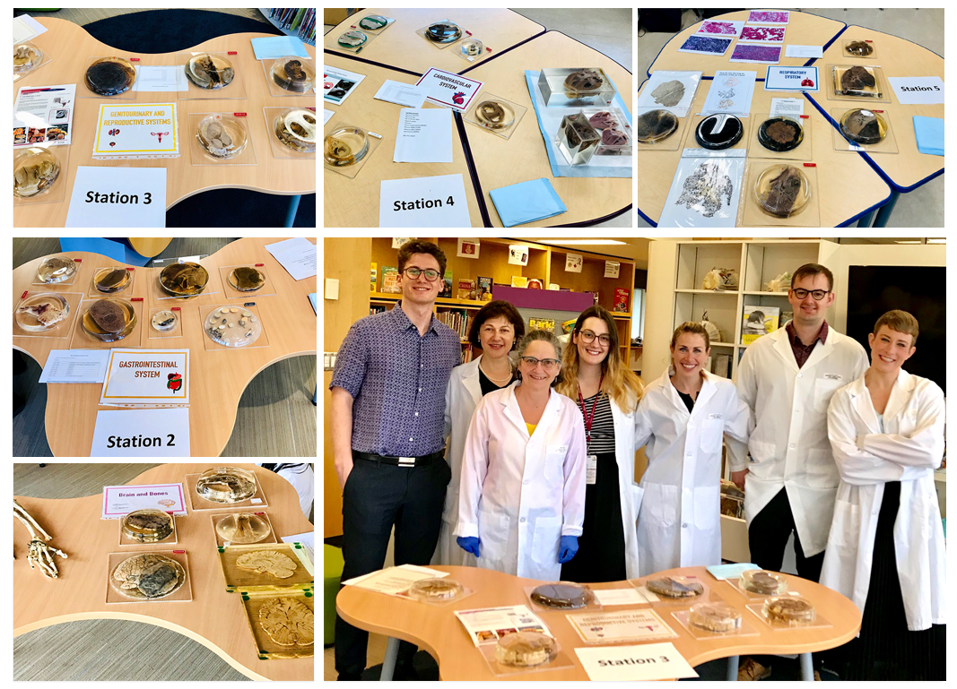
When Dr. Ionescu asked me if I would like to join her and help on an education session called “The Inside of Us”, I didn’t really know what to expect, but was really intrigued by the opportunity. Dr. Ionescu assembled a team consisting of pathology residents Mike Steel, Kimberly Hamilton, Lisa Borretta, and Helen Dyck. At the invitation of the grade 6th science teacher, Ms. Stephanie Browning, the team presented an anatomy and pathology lesson to 75 students, of West Point Grey Academy (WPGA), where Dr. Ionescu’ son Andrew is studying. As we entered WPGA, we were welcomed by the smiling faces of the teachers, as well as the glimmer of the middle school’s “STEM lab”, full of technology gadgets. As we began to unload boxes with over 50 specimens of human organs from Dr. Ionescu’s car, the excitement began. The specimens were carefully selected from the William Boyd Museum, and categorized by organ system, with the invaluable help and expertise of Mrs. Helen Dyck, the Manager & Curator of the museum. As part of this session, specimens were set up in separate stations where students were able to handle them. There were 5 stations, comprising of the cardiovascular, respiratory, genitourinary and reproductive, and gastrointestinal systems, as well as a brain and bone station.
Each station was run by a member of the team wearing a lab coat (for scholarly effect), describing the specimens, and answering questions. After a quick introduction by Dr. Ionescu, 5 groups of 15 kids rotated between the stations for mini interactive lectures. Some students were bewildered by what they saw, some merely floated, and a few needed to grab some water. This had been the first group of children younger than grade 8 to observe these specimens!
Each group was assigned a teacher who followed the children, googly-eyed, “making sure the children were respectful”. Asking questions left and right, most of the adults had gone their whole lives without seeing anything like this unique experience! Looking back, I am not sure who were more impressed - the teachers or the children.
The respiratory system station was a big hit. Normal lungs, black emphysematous lungs from a heavy smoker, lung cancer, and a lung of a toddler with a peanut stuck in one of the bronchi were displayed.
The respiratory system station was a big hit. Normal lungs, black emphysematous lungs from a heavy smoker, lung cancer, and a lung of a toddler with a peanut stuck in one of the bronchi were displayed. The children asked many questions about the effects of smoking and were astounded to learn that even healthy lungs of non-smokers have black spots due to environmental pollution. Later on, we received feedback and thanks from a mom saying that her son “would never smoke, but after seeing the smokers’ lungs he won’t let anyone smoke either” -- pretty powerful.
The children, bewildered by what they had just experienced, were shortly after off to their next class and so was I… on to my next pathology adventure.
After the 5 rotations and 90 minutes had passed, the presenters were exhausted from the energy of the children picking their brains. With a gracious thank you from Ms. Browning, the WPGA experience concluded. The children, bewildered by what they had just experienced, were off to their next class and so was I… on to my next pathology adventure.
Forensic Pathology at the Morgue:
I, on the other hand, was simply trying to determine whether or not what I was seeing was real, and reminding myself to breathe through my mouth so I wouldn’t faint!
Fast forward a month, and I’m in the basement of Vancouver General Hospital (VGH) putting on scrubs for my first ever visit to a morgue. As I entered the post-mortem suite, there were three bodies lying on stainless steel tables with large scales dangling above them. A group of pathologists were gathered around one of the bodies, carefully inspecting and gently palpating in their search for signs of disease and injury. I, on the other hand, was simply trying to determine whether or not what I was seeing was real, and reminding myself to breathe through my mouth so wouldn’t faint!
Dr. Orde then wandered over to his computer and queued up Queen onto the speakers, which echoed through the morgue and quickly livened things up. Teams of pathologists and their assistants commenced their examination of the bodies’ internal structures, and as I started looking at the bodies through the lens of science, it dawned upon me that this might just be one of the best ways to learn anatomy. I watched as a resident took out the entire abdominal contents of an individual, but when we got to the brain I was asked to help out! Full of excitement, under the supervision of Dr. Orde, we very diligently removed the brain with meticulous guidance after the deceased skull was carefully sawed open. In the background I heard the familiar bass riff of “Another One Bites the Dust”, but nobody else seemed to pay attention to the irony of this song blaring through the morgue.
Overall, this day was a success, being both extremely informative and interesting. It was fascinating to see how patient care continues even after death, and to discover that the autopsy is a key component of healthcare, as well as providing much needed answers for family members, coroners, and the public at large. After the autopsies, off I went with a pen in hand that had the label “our days begin when your days end”. Thank you Dr. Orde and all at VGH morgue for making this possible! I can’t wait to see what next year has to bring!